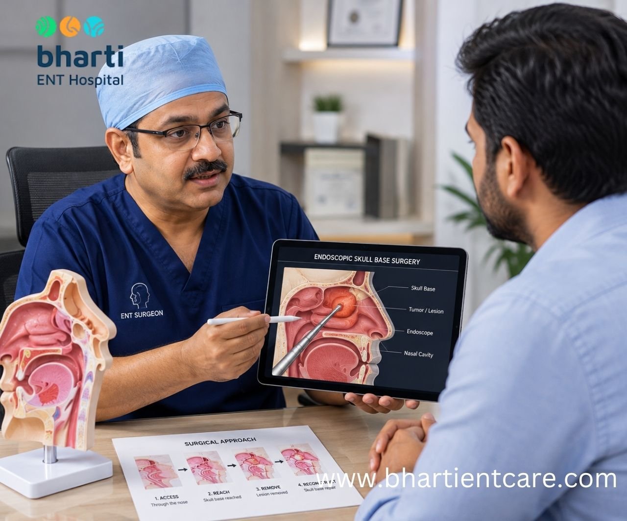
Endoscopic skull base surgery is a surgical procedure performed with a small camera known as an endoscope (used sometimes through the nose) to access specific areas of the skull base without a large incision into the scalp or skull.
Skull base is the bottom of the skull which lies between the brain and the structures of the face, nose and upper airway. It is in close proximity or contains significant structures such as the pituitary gland, major blood vessels, nerves and areas of vision and smell. Due to this complicated anatomy, careful planning and special experience in surgery is required in this region.
In an endoscopic endonasal procedure, the surgeon can access specific lesions at the skull base through the natural endonasal (nose) cavity and sinuses. The technique has been widely utilized in pituitary surgery and has since been extended to certain skull base diseases.
The technique is sometimes referred to as “minimally invasive” because it doesn’t involve some of the outside incisions, removal of facial bone and brain retraction that are used with other methods. But small incisions does not equate to no risk or all skull base surgeries.
Why Is the Skull Base Such an Important Area?
The skull base is important because it forms a boundary between the brain and the structures of the face and contains several critical nerves, blood vessels and organs.
It is not simply a layer of bone. Different areas of the skull base are located close to structures responsible for vision, hormone regulation, facial movement, hearing, smell and other neurological functions.
Some lesions can therefore be difficult to access safely using conventional surgery.
The exact surgical route depends on factors such as:
- Location of the lesion
- Size of the lesion
- Direction in which it extends
- Relationship with major blood vessels
- Relationship with nerves
- Involvement of the pituitary gland
- Involvement of the optic nerves
- Whether the lesion extends toward the nasal cavity or sinuses
- Whether there is an existing or expected CSF leak
This is why skull base surgery is highly individualized. Two patients with apparently similar diagnoses may not necessarily require the same surgical approach.
How Is Endoscopic Skull Base Surgery Performed?
In most cases, a surgeon will approach targeted sections of the skull base in an endoscopic endonasal skull base surgery via the nose with an instrument called an endoscope. The specific approach will depend on the location and nature of the disease.
The endoscope has a camera and light source to enable the surgical team to see the anatomy on a high-definition monitor.
Instead of making a big opening on the outside, the surgeon may enter via the nose with appropriately selected lesions.
Depending on the condition, the operation may involve several stages:
1. Preoperative imaging and planning
CT or MRI scans can help to visualise the anatomy and determine the location and size of the lesion.
Detailed imaging is especially important, as the surgeon must understand the relationship of the lesion with adjacent nerves and blood vessels, the pituitary gland and other structures.
2. Endoscopic access through the nose
The surgical team introduces the endoscope through the nasal cavity and creates the appropriate surgical corridor.
The route depends on where the lesion is located.
3. Reaching the skull base
The surgeon carefully works through the relevant nasal or sinus structures to reach the skull base.
Different approaches can be used for different anatomical regions.
4. Treating or removing the lesion
The surgeon uses specialized instruments while viewing the operative field through the endoscope.
The goal may be complete removal, decompression, biopsy or another disease-specific treatment, depending on the diagnosis.
5. Skull base reconstruction
However, if a defect is left after surgery where the CSF could leak, the base of the skull may need to be reconstructed.
Depending on the size and nature of the defect and the possibility of CSF leakage, there are different reconstruction techniques that can be applied, such as graft, sealants and vascularized tissue flaps.
This reconstruction step is crucial to the endoscopic skull base surgery and not a mere optional last step.
When Is Endoscopic Skull Base Surgery Recommended?
Endoscopic skull base surgery is reserved for specific conditions where the site, size and morphology of the lesion allows for an endoscopic approach and where it is safe.
Not all skull base tumors/lesions will receive this treatment.
Endoscopic endonasal surgery has been applied to selected tumors of the midline skull base, such as pituitary tumors and some other skull base tumors. Applications include conditions like craniopharyngiomas, some meningiomas and chordomas, but it is ideal for each patient.
Depending on the patient’s diagnosis, an endoscopic approach may be considered for:
- Selected pituitary tumors
- Certain anterior skull base tumors
- Selected skull base tumors extending toward the nasal or sinus region
- Some CSF leaks
- Certain lesions involving the clivus or midline skull base
- Selected conditions affecting the sellar or parasellar region
- Certain skull base defects
The decision should be based on imaging, diagnosis, anatomy and the experience of the surgical team.
Can Pituitary Tumors Be Removed Through the Nose?
Yes. Some pituitary tumors can be treated with an endoscopic endonasal or transsphenoidal surgery via the nose.
This is one of the most popular endoscopic skull base surgeries.
The pituitary is housed in a bony pocket called the sella turcica, which is close to the middle of the base of the skull. The position makes it possible to access the pituitary area through the nose and sphenoid sinus in some patients.
Endoscopic techniques have increased access to the pituitary region and other areas around the midline of the skull base without exposing as much as the traditional transcranial surgery.
But not all pituitary tumors can be best treated in the same way.
The surgical approach may be influenced by the size of the tumour, the extent of spread, involvement of veins, involvement of the optic apparatus and involvement of the cavernous sinus.
So, the question isn’t whether or not a pituitary tumor can be removed via the nose.
The key question is:
Do you think this particular pituitary tumour is suitable for the endoscopic endonasal approach?
What Other Conditions Can Be Treated With Endoscopic Skull Base Surgery?
Selected skull base tumors, CSF leaks and lesions located around the midline skull base may be treated using endoscopic approaches when their anatomy makes this route appropriate.
The range of conditions treated through endoscopic skull base approaches has expanded significantly since the technique was first used primarily for pituitary surgery.
Depending on the location and diagnosis, examples can include:
Pituitary tumors
Endoscopic transsphenoidal surgery is an established approach for selected pituitary tumors.
Selected skull base tumors
Some tumors involving the anterior skull base, clivus or other midline areas may be accessible through an endoscopic endonasal route.
CSF leaks
Some cerebrospinal fluid leaks involving the skull base can be repaired endoscopically through the nose.
Selected meningiomas and craniopharyngiomas
Certain lesions in anatomically suitable locations may be considered for an endoscopic approach, although treatment decisions are highly individualized.
The key point is that diagnosis alone does not determine the surgical route. Location and anatomy are equally important.
What Is a CSF Leak and Why Does It Matter in Skull Base Surgery?
The CSF leak is a condition where cerebrospinal fluid leaks out because of a defect in the protective layers around the brain and preventing or repairing the CSF leak is an important aspect of skull base surgery.
The brain and spinal cord are surrounded and protected by cerebrospinal fluid (CSF).
In certain skull base surgeries, a space between the brain and the nose/sinus cavity may be opened. If the opening is not properly reconstructed, CSF can drain into the nose.
Hence, skull base reconstruction plays an important role in endoscopic skull base surgery.
Leakage of CSF can make complications more likely, such as infection (meningitis). There are surgical techniques that have been developed that have helped minimize CSF leakage rates, such as the vascularized nasoseptal flaps in certain patients.
The reconstruction technique is selected according to factors such as:
- Size of the skull base defect
- Location of the defect
- Presence and amount of CSF flow
- Surgical approach used
- Patient-specific anatomy
- Intraoperative findings
This is one reason why skull base surgery requires detailed preoperative planning and specialized intraoperative decision-making.
Is Endoscopic Skull Base Surgery Minimally Invasive?
Endoscopic skull base surgery is considered minimally invasive compared with some traditional open approaches because it can use the nasal corridor and avoid certain external incisions, facial bone flaps and brain retraction.
However, “minimally invasive” should not be interpreted as “simple.”
The skull base contains highly sensitive structures, and surgery can involve technically demanding anatomy.
The potential advantages of an endoscopic endonasal approach may include:
- No large scalp incision for appropriately selected procedures
- Avoidance of some forms of craniotomy
- Direct access to selected midline skull base lesions
- Magnified endoscopic visualization
- Potentially less disruption of some surrounding structures
- No external facial scar from a traditional craniofacial incision
Research reviews have described the endonasal endoscopic approach as an important option for selected skull base pathologies, while also emphasizing that the technique still has significant potential complications and limitations.
Therefore, the correct question is not whether endoscopic surgery is always better.
It is whether endoscopic surgery is the best approach for the patient’s particular anatomy and disease.
Is Endoscopic Skull Base Surgery Safe?
Endoscopic skull base surgery can be performed safely in appropriately selected patients, but it is a complex procedure with potential complications that must be discussed before surgery.
Like other major surgeries, the risks depend on the disease, surgical approach, anatomy and experience of the surgical team.
Potential complications discussed in the medical literature include:
- Cerebrospinal fluid leak
- Infection
- Pituitary or hormonal dysfunction
- Vision-related complications
- Cranial nerve dysfunction
- Blood vessel injury
- Nasal or sinus-related problems
- Bleeding
- Need for additional treatment or surgery
The risk profile is not identical for every procedure.
For example, a straightforward endoscopic pituitary operation and an expanded skull base procedure involving structures beyond the sella can have very different technical requirements.
Published reviews emphasize CSF leakage, cranial nerve dysfunction, pituitary dysfunction, vascular injury and infection among recognized complications of endoscopic endonasal approaches.
This is why patients should discuss the specific risks of their own procedure with their surgical team rather than relying on a general complication rate found online.
How Do Doctors Decide Whether a Patient Is Suitable for Endoscopic Skull Base Surgery?
Doctors determine suitability by evaluating the diagnosis, imaging, tumor location, size, extension and relationship with nearby structures, along with the patient’s overall health and the expertise available within the treating team.
A skull base operation generally requires much more planning than simply deciding whether a tumor can be reached with an endoscope.
Doctors may review:
- MRI findings
- CT scan findings
- Tumor size
- Tumor location
- Extension toward surrounding structures
- Relationship with the optic nerves
- Relationship with major blood vessels
- Pituitary function
- Nasal and sinus anatomy
- Previous surgery
- Overall medical condition
In complex cases, the decision may involve more than one specialty.
The modern skull base approach often relies on collaboration between ENT/skull base surgeons, neurosurgeons, radiologists, endocrinologists, ophthalmologists or other specialists, depending on the patient’s condition.
Recent literature on expanded endoscopic approaches emphasizes the importance of multidisciplinary teams and experience when treating technically complex skull base tumors.
Should the neurosurgeon be involved in Endoscopic Skull Base Surgery?
The team composition differs for each endoscopic skill base procedure, but sometimes it may be necessary for an ENT/skull base surgeon to work closely with the neurosurgeon in a complex skull base surgery.
The base of the skull is a junction of several anatomical areas.
An ENT/skull base surgeon will have experience of the nasal cavity, sinuses, endoscopic access and reconstruction, while a neurosurgeon might be responsible for the intracranial aspect of the disease.
Surgery will require a specific team, depending on the diagnosis and surgery.
This type of collaboration is especially significant when it comes to complex lesions that affect the brain, pituitary area, large blood vessels or large defects in the base of the skull.
So, it is important for patients to check for the medical centre’s specialization and facilities to treat the skull base condition and not just any ENT consultation in Vadodara.
What Tests Are Done Before Endoscopic Skull Base Surgery?
Preoperative testing is used to understand the lesion, map the anatomy and assess the patient’s suitability for surgery.
Imaging is a major part of the planning process.
Depending on the condition, doctors may request:
- MRI of the brain and skull base
- CT scan of the nose, sinuses and skull base
- Hormonal testing for pituitary conditions
- Vision or ophthalmological assessment
- Nasal endoscopic examination
- Routine preoperative blood investigations
- Other condition-specific tests
The purpose of imaging is not only to locate the lesion.
It helps the surgical team understand the three-dimensional relationship between the lesion and critical anatomical structures.
For complex skull base surgery, detailed imaging can also help determine the anticipated surgical corridor and reconstruction strategy.
What to expect during Skull Base Reconstruction?
Once the lesion has been removed or a hole has been made in the base of the skull, the surgeon can fill in the hole with a suitable graft, sealants or vascularized tissue to aid in the restoration of the barrier between the nasal cavity and the cranial vault.
Reconstruction is very critical when there is the possibility of CSF leakage.
The common technique being discussed is the vascularized nasoseptal flap which utilizes the tissue from the nasal septum to cover an appropriate defect in the skull base.
Vascularized nasoseptal flap reconstruction after ETSS has been demonstrated to be beneficial in preventing CSF leakage following surgery in comparison to some nonvascularized techniques.
But not all patients will need the same reconstruction.
The approach relies on the defect size and location, CSF flow and in the operating room.
For this reason the reconstruction should not be the afterthought but a part of skull base surgery.
What are the benefits of skull base surgery, endoscopically?
The endonasal approach to the skull base may directly access some lesions of the skull base without the disruption of some traditional open approaches, although it is not appropriate for all patients.
Potential advantages include:
- Access via the natural nasal passage
- The replacement of a large external scalp incision in many procedures
- A procedure for directly visualizing with an endoscope.
- Access to selected areas of the midline skull base
- With the correct patient selection, the use of a retractor will be diminished.
- May have a shorter recovery period than some open methods
However, the benefits are very much related to the patient’s condition.
Not all endoscopic procedures are better than open surgery. Some lesions are located in areas where an open or alternative surgical approach may be safer and/or more effective.
The objective should always be to treat the person as safely and effectively as possible and not just because the method is more recent or less invasive.
So, What are the Limitations of Endoscopic Skull Base Surgery?
Not all skull base tumours and conditions can be treated by endoscopic surgery and certain of the tumours may require an open or alternative surgical technique.
Skull base anatomy is very variable.
An endoscopic approach is not optimal due to:
- Tumor location
- Lateral extension
- Major blood vessels are involved.
- The process of involvement of critical nerves.
- Extensive intracranial extension
- Previous surgery
Anatomical variations in the nose or sinuses, which may affect a patient’s nasal or sinus passages.
The use of a different surgical angle is required.
That is why patients should check with caution the statements that the endoscopic surgery is always “safer,” “scarless” or “better.
The choice of approach should be made after examining the imaging and diagnosis of the patient.
What is the duration of Endoscopic Skull Based Surgery procedure?
The duration of endoscopic surgery of the skull base is variable and cannot be standardized, depending on the condition, surgical approaches, tumor size and complexity.
A relatively simple endoscopic pituitary procedure can be completed in a very different time than a long-duration skull base tumor surgery which needs reconstruction.
Operating time can be influenced by the following factors:
- Size of the lesion
- Location
- Surgical approach
- The extent of removal necessary
- Presence of CSF leakage
- Reconstruction requirements
- Previous surgery
The complexity of surrounding anatomy is a factor.The surrounding anatomy is complex.
Patients should therefore check with their surgical team to determine a time frame for their procedure, as opposed to using an online average.
What to expect after Endoscopic Skull Base Surgery?
The recovery depends on the level and type of surgery performed and the patient’s health and whether complications occurred or further surgery was performed.
Recovery isn’t a one-sided thing as endoscopic skull base surgery may require recovery from both the surgical wound and the underlying condition.
Patients may require follow up to monitor:
- Nasal healing
- Skull base reconstruction
- CSF leakage
- Vision
- Hormonal function if pituitary is affected
- Infection
- Symptoms related to the original disease
The specific limitations and post surgical program should be outlined by the surgical team.
Patients should also be aware that recovery time is not the same as the time until the final outcome of patients’ treatment. After surgery some tumors need to be pathologically reviewed, monitored with hormones or followed up with a radiologic exam.
The Warning Signs for Patients After Skull Base Surgery?
There are potential complications that can occur after surgery and patients should seek immediate surgical attention if they develop symptoms suggestive of CSF leakage, infection, significant bleeding, worsening neurological symptoms, or any other unexpected changes.
Some symptoms that could be concerning are:
- Clear watery drainage from the nose
- Persistent severe headache
- Fever
- Neck stiffness
- Significant nasal bleeding
- Worsening vision
- New neurological symptoms
- Persistent vomiting
Worsening or worsening pain.
After skull base surgery, if the nasal discharge is clear and watery, it does not necessarily mean it is normal nasal mucus. Patients are advised to get in touch with their health care team to evaluate the symptom.
In the event of severe neurological symptoms, breathing difficulties or emergencies, prompt medical care is suitable.
Is it possible to do Endoscopic Skull Base Surgery without any scar on the face?
The nasal cavity is the surgical route for many endoscopic endonasal skull base procedures and thus there is no need for a large external facial incision.
This is one of the reasons why it is appealing for lesions that are specifically chosen.
The surgeon is able to enter the body through the nostril and nasal cavity with the aid of an endoscope and specialized instruments.
This should not, however, be interpreted to mean that there are no tissues that are injured by the procedure, or that recovery is always effortless.
There is also a fair amount of surgery performed on the nasal tissues and the base of the skull, which may cause temporary or permanent nasal problems.
Surgical treatment should therefore be planned for treatment with the treating surgeon in view of the diagnosis.
Does there exist Endoscopic Skull Base Surgery at Vadodara?
Yes. Some of the other services offered by Bharti ENT Care are Skull Base Surgery, Anterior Skull Base Surgery and CSF Leak Repair which are all related to the field of ENT.
The hospital, in particular, specifies:
- Skull Base Surgery
- Anterior Skull Base Surgery
- CSF Leak Repair
- Endoscopic Sinus Surgery
One of its ENT services is among them.
Bharti ENT Care further claims to have an operation theatre set up for skull base and endoscopic ENT surgery and has an equip named Karl Storz high-definition endoscopic camera system.
For patients coming from Vadodara and nearby areas, this implies the diagnostic and imaging workup can start with a detailed assessment of the diagnosis prior to deciding on the suitability of using an endoscopic skull base approach.
What is the approach of Bharti ENT Care in the field of Skull Base Surgery?
Bharti ENT Care claims to provide diagnosis, treatment, and postoperative care; the centre is equipped with advanced surgical equipment and special services offered by the ENT department.
The hospital says its model is “consultation, investigations, surgery and care after surgery are all in one place. It also claims physicians listen to patients’ health issues and offer one-on-one counseling.
The process would typically require individualization based on patient’s underlying condition when the patient is being evaluated for skull base surgery.
The assessment may involve:
Understanding the diagnosis
The doctor examines the patient’s symptoms and the patient’s prior treatment and confirmed or suspected diagnosis.
Reviewing imaging
MRI and CT imaging can be used to help identify the location and size of the lesion and how it is related to other tissue and structures.
Identifying the operating approach
The team assesses if an endoscopic endonasal approach is suitable and if there is an alternative surgical pathway which will allow safer access.
Planning reconstruction
If the procedure causes a defect in the skull base or is likely to leave a CSF leak, then planning for reconstruction is an important component of the procedure. Depending on the case, modern skull base reconstruction can be multi-layer and with vascularized tissue flaps.
Providing postoperative follow-up
Follow up is essential to provide reassurance regarding treatment outcome, complications and healing.
Skull base surgery can also be very complicated, and patients should also inquire if they will need to work with neurosurgery or another specialty for their specific case.
Frequently Asked Questions About Endoscopic Skull Base Surgery
What is endoscopic skull base surgery?
Endoscopic skull base surgery is a relatively new minimally invasive surgical technique that accesses selected lesions in the skull base via an endoscope, typically through the nose. Used for conditions that are correctly chosen, depending on anatomical location and disease.
Does endoscopic skull base surgery involve nasal surgery?
The nose is the entry point for many endoscopic methods of surgical approaches to the base of the skull. The endonasal approach is a good option for accessing certain areas of the mid-skull base without the extensive external incisions of some other surgical methods.
Is it possible to remove a pituitary tumour by the nose?
Yes. Some pituitary tumors may be treated with an endoscopic endonasal or transsphenoidal procedure. This is suitable or not according to the size of the tumor, its location, its extension, and the patient’s anatomy.
Will endoscopic skull base surgery hurt?
It is a complex procedure and can be performed safely in appropriately selected patients, but carries some risk. Possible complications include CSF leak, infection, hormonal dysfunction, cranial nerve problems, and vascular injury and others depending upon the surgery performed.
Which conditions can be treated using endoscopic skull base surgery?
Some CSF leaks and other appropriately located lesions as well as selected pituitary tumours can be treated endoscopically. Indication will depend upon the imaging and the diagnosis of the patient.
Is there any scar after endoscopic skull base surgery?
Most endonasal surgeries are not surgically performed in the face or scalp, but rather through an endonasal approach. The surgery does however require manipulation of tissue within the nose and skull base.
A CSF leak following skull base surgery is a common complication.
A CSF leak is when CSF can leak out through a hole in the protective layers around the brain. Skull base reconstruction is performed when required to minimize the chances of CSF leakage in the postoperative period.
What is the recovery period post endoscopic skull base surgery?
Recovery depends on the type and complexity of surgery as well as the condition of the patient. Follow-up may involve checking the nose for healing, CSF leakage, vision, hormonal function and the underlying disease.
Is Endoscopic skull base surgery superior to open skull base surgery?
Not necessarily. For certain lesions, endoscopic surgery may provide valuable benefits, but the best way to approach the surgery depends on the location, extent and size of the condition. Other lesions are more appropriate to open or alternative treatments.
Where to seek guidance for a Skull base surgery in Vadodara?
The services offered by Bharti ENT Care, Vadodara include: Skull Base Surgery, Anterior Skull Base Surgery and CSF Leak Repair.